610 Quintard Avenue, Oxford, AL 36203 | Phone: (256) 831-6116 | Fax: (866) 928-5017 | Mon-Fri 9:00am - 7:00pm | Sat 9:00am - 6:00pm | Sun 12:00pm - 6:00pm
Get Healthy!
Recent health news and videos.
Staying informed is also a great way to stay healthy. Keep up-to-date with all the latest health news here.
14 Jul
Obesity-Related Cancer Deaths Tripled Over the Past Two Decades, Study Finds
Researchers discover steep increases in cancer deaths linked to obesity since 1999, especially among women, older adults and Black people.
11 Jul
‘Alarming Rise’ in Gastrointestinal Cancers in Young Adults
A new study finds early-onset gastrointestinal cancers increased by nearly 15% from 2010 to 2019.
10 Jul
Severe Withdrawal From Antidepressants Uncommon, Study Finds
A review of 50 clinical trials on antidepressants and withdrawal concludes most patients experience one additional symptom, the most common being dizziness and nausea.

CDC Says COVID-19 Cases Rise in 25 States
COVID-19 cases are on the rise again across the United States, with the biggest increases in parts of the South, Southeast and West Coast.
The U.S. Centers for Disease Control and Prevention (CDC) estimates that 25 states are seeing growth in COVID cases as a summer wave appears to be starting, CBS News reported.
Even though...
- I. Edwards HealthDay Reporter
- |
- July 14, 2025
- |
- Full Page

Bread Sold at Walmart, Kroger Recalled for Hazelnut Allergy
A popular brand of bread sold at Walmart, Kroger and other stores has been recalled in 12 U.S. states due to undeclared hazelnuts.
Hartford Bakery Inc. is recalling six lots of its Lewis Bake Shop Artisan Style 1/2 Loaf bread because they may contain hazelnuts, a tree nut that can trigger dangerous allergic reactions, People repor...
- I. Edwards HealthDay Reporter
- |
- July 14, 2025
- |
- Full Page
Arizona Resident Dies From Pneumonic Plague
An Arizona resident has died from pneumonic plague, a rare but serious illness, health officials confirmed.
The death happened in Coconino County in Northern Arizona, which includes Flagstaff. Officials gave no information about the person who died, The Associated Press reported.
This is the first known U.S. death from...
- I. Edwards HealthDay Reporter
- |
- July 14, 2025
- |
- Full Page

Diet Influences Constipation Risk, Study Says
Worried about maintaining your regularity as you grow older?
Changing your diet can reduce the risk of chronic constipation in middle-aged folks and seniors, a new study says.
The Mediterranean diet and plant-based diets were found to best ward off constipation, researchers reported recently in the journal Gastroenterology.<...
- Dennis Thompson HealthDay Reporter
- |
- July 14, 2025
- |
- Full Page

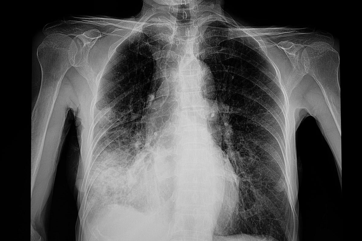
Household Mold Can Trigger Lung Disease
Household mold can be a significant trigger for a rare but potentially debilitating lung disease that can cause permanent breathing problems, a new study says.
Mold appears to be the primary cause for nearly a quarter (23%) of 231 cases of hypersensitivity pneumonitis (HP) treated at the University of Texas Southwestern Medical Center in D...
- Dennis Thompson HealthDay Reporter
- |
- July 14, 2025
- |
- Full Page

New Bride Survived Removal Of A 40-Pound Tumor
A young woman became a June bride months after surviving complex surgery to remove a 40-pound tumor from her body, Cedars-Sinai doctors report.
Adriana Pulido, 24, died on the operating table six times during an early attempt to remove the huge tumor.
Located near her liver, the tumor had grown so large that it had displaced her hear...
- Dennis Thompson HealthDay Reporter
- |
- July 14, 2025
- |
- Full Page

Sugar, Sweeteners Might Trigger Early Puberty In Some Kids
Sugar and artificial sweeteners might increase the risk of early puberty in children, a new study says.
Sugar, aspartame (Equal), sucralose (Splenda) and glycyrrhizin (licorice root) are all significantly associated with a higher risk of early puberty, particularly in genetically predisposed children, researchers reported Sunday at the End...
- Dennis Thompson HealthDay Reporter
- |
- July 14, 2025
- |
- Full Page

Menopause Hormone Therapy Boosts GLP-1 Drug Effectiveness, Researchers Say
Good news for women of a certain age: Hormone replacement therapy for menopause appears to boost the effectiveness of GLP-1 weight loss drugs like Wegovy and Zepbound, a new study says.
Women using tirzepatide (Zepbound) lost more weight if they also were taking hormone therapy — 17% of body weight lost versus 14% after a median 18 m...
- Dennis Thompson HealthDay Reporter
- |
- July 14, 2025
- |
- Full Page

Obesity-Related Cancer Deaths More Than Triple In U.S.
Cancer deaths linked to obesity more than tripled in the U.S. during the past two decades, a new study says.
Deaths linked to the 13 types of obesity-related cancer rose to 13.5 deaths per million from 3.7 deaths per million between 1999 and 2020, researchers reported Sunday at the Endocrine Society’s annual meeting in San Francisco....
- Dennis Thompson HealthDay Reporter
- |
- July 14, 2025
- |
- Full Page

How to Stay Safe at the Beach or Pool This Summer
A day at the beach or pool is a great way to beat the summer heat — but it can also be risky if you’re not careful.
Dr. Eric Costanzo of Hackensack Meridian Jersey Shore University Medical Center has some valuable advice to help families stay safe this summer, whether at the shore or at home by the pool.
The ocean c...
- I. Edwards HealthDay Reporter
- |
- July 13, 2025
- |
- Full Page

Killer Whales Show a Softer Side, Sharing Food With People
They’re known as "killer whales," but orcas have a surprisingly soft and even generous side.
"Orcas often share food with each other — it’s a prosocial activity and a way that they build relationships with each other. That they also share with humans may show their interest in relating to us as well," said Jared Towers, l...
- Carole Tanzer Miller HealthDay Reporter
- |
- July 12, 2025
- |
- Full Page

About 1 in 3 U.S. Teens Now Have Prediabetes, New CDC Data Shows
A new government estimate shows that nearly 1 in 3 U.S. teens have prediabetes, putting them at risk for Type 2 diabetes and other serious health problems.
In a new analysis, the U.S. Centers for Disease Control and Prevention (CDC) says about 8.4 million 12- to 17-year-olds were prediabetic in 2023. That’s far more than the 18% esti...
- I. Edwards HealthDay Reporter
- |
- July 11, 2025
- |
- Full Page
RFK Jr. Cancels Key U.S. Health Panel Meeting Without Warning, Raising Concerns
U.S. Health Secretary Robert F. Kennedy Jr. canceled a meeting of government health panel that helps guide what preventive care is covered by insurance, alarming doctors and other health officials.
The U.S. Preventive Services Task Force (USPSTF) was set to meet Thursday, but members were told in an email Monday that the meeting would be p...
- I. Edwards HealthDay Reporter
- |
- July 11, 2025
- |
- Full Page

Ritz Peanut Butter Crackers Recalled Over Labeling Mistake
A labeling mistake has led to a nationwide recall of four different types of Ritz Peanut Butter Cracker Sandwich cartons, federal health officials said.
Mondelēz Global LLC, the parent company of Ritz, is recalling the products because some packages containing peanut butter may be incorrectly labeled as cheese, the U.S. Food and Drug ...
- I. Edwards HealthDay Reporter
- |
- July 11, 2025
- |
- Full Page

FDA Fully Approves Moderna’s COVID Vaccine for Some Young Kids
Moderna’s COVID-19 vaccine has received full approval from the U.S. Food and Drug Administration (FDA) for use in children with medical conditions that put them at higher risk of severe illness.
The move makes Moderna’s vaccine, called Spikevax, the first COVID shot for kids in the U.S. to be fully approved, rather than used un...
- I. Edwards HealthDay Reporter
- |
- July 11, 2025
- |
- Full Page

U.S. Cuts to HIV Aid Could Lead to 4 Million Deaths, U.N. Warns
Global deaths from AIDS have dropped to their lowest levels in more than 30 years, in part due to efforts to fight HIV. But U.S. funding cuts could soon reverse that progress, experts warn.
A United Nations report released Thursday says that if the money isn’t replaced, more than 4 million people could die of AIDS by 2029 and 6 milli...
- I. Edwards HealthDay Reporter
- |
- July 11, 2025
- |
- Full Page

Antidepressant Withdrawal Not As Severe As Thought, Evidence Review Says
People typically don’t suffer severe withdrawal symptoms or fall into depression immediately after they stop taking antidepressants, a new evidence review says.
There had been concerns that people who quit antidepressants would suddenly fall prey to depression or develop what’s known as antidepressant discontinuation syndrome.<...
- Dennis Thompson HealthDay Reporter
- |
- July 11, 2025
- |
- Full Page

Popular Chronic Pain Med Linked To Dementia Risk
A drug used to treat seizures, nerve pain and restless leg syndrome might be linked with increased risk of dementia, a new study says.
Regular gabapentin use appeared to increase risk of dementia by 29% and mild cognitive impairment (MCI) by 85%, researchers reported July 10 in the journal Regional Anesthesia & Pain Medicine.<...
- Dennis Thompson HealthDay Reporter
- |
- July 11, 2025
- |
- Full Page

Societal Stress Driving Trans, Gender-Diverse To Drink
Societal stress could be driving some transgender and gender-diverse people to the bottle, a small-scale study says.
People whose gender identity differs from their birth sex drink more alcohol and engage in more hazardous drinking than cisgender heterosexual peers, according to findings published July 6 in the journal Alcohol: Clinica...
- Dennis Thompson HealthDay Reporter
- |
- July 11, 2025
- |
- Full Page

Disposable Vapes Release Toxic Metals, Lab Study Says
People using cheap disposable vape devices are likely inhaling high levels of toxic metals with every puff, a recent study says.
After a few hundred puffs, some disposable vapes start releasing levels of toxic metals higher than found in either last-generation refillable e-cigarettes or traditional tobacco smokes, researchers reported in t...
- Dennis Thompson HealthDay Reporter
- |
- July 11, 2025
- |
- Full Page
